Dental X Ray Radiation Dose Chart
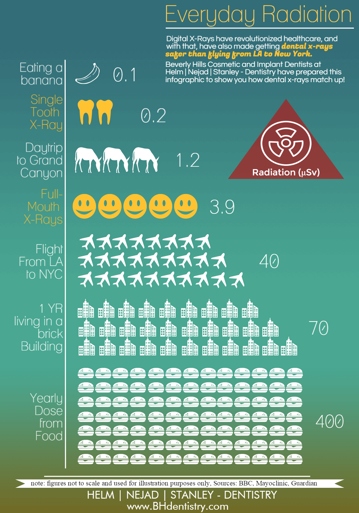
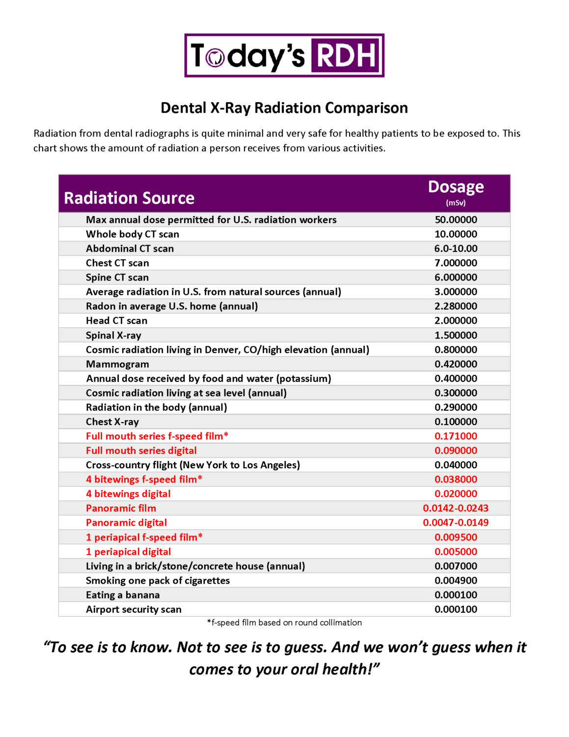
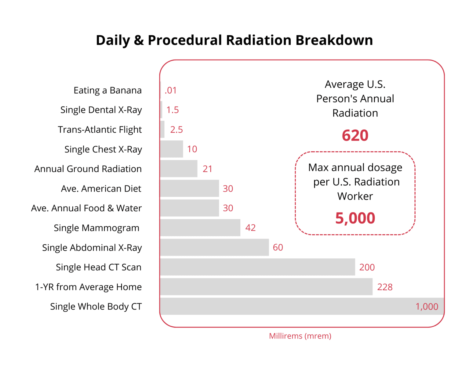
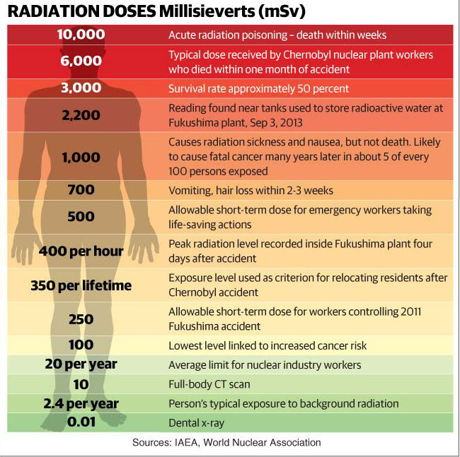
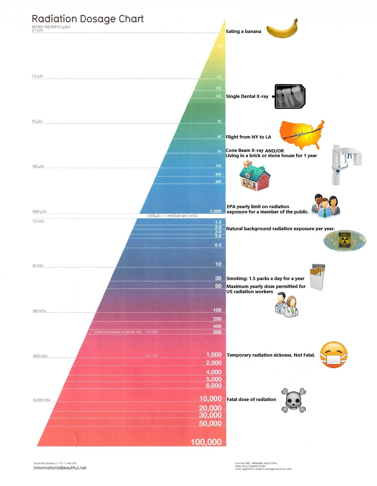
Dental X Ray Radiation Dose Chart - Dental radiographs can alert your dentist to changes in your hard and soft tissues. Web normal daily background dose for an average person: This chart shows the amount of radiation a person receives from various activities. Radiation workers 50.00000 whole body ct scan 10.00000 An expert panel presents recommendations on radiation safety, appropriate imaging practices, and reducing radiation exposure. Web in figure 1, the dose from two intraoral films is 0.002 msv. Web because of the low radiation dose associated with dental radiographs, people who have received radiation treatment for head and neck cancer can undergo dental radiography safely. ─ some types of tumors. Airplane flight from ny to los angeles: Web comparison of the total effective dose (effective dose multiplied by the number of image sets) with that from other radiation sources: ─ infections that develop under your gums; Web because of the low radiation dose associated with dental radiographs, people who have received radiation treatment for head and neck cancer can undergo dental radiography safely. Radiation source dosage (msv) max annual dose permitted for u.s. ─ some types of tumors. Web here are some approximate comparisons of background radiation and effective radiation dose in adults for several radiology procedures described on this website. Radiation workers 50 whole body. Web • going through an airport security scanner 80 times is the equivalent to a single day of casual radiation exposure. Yearly cosmic radiation while living at sea level: Web effective dose calculations based on the revised guidelines given by the international commission on radiological protection (icrp 103). Web typical effective doses are for: This chart shows the amount of radiation a person receives from various activities. For occlusal radiographs, the dap was 7.43 cgy × cm 2 and the ed 2.22 µsv. Overall, 70.2% of all intraoral radiographs were dental, 20.3% bitewing and 9.5% occlusal radiographs. Web normal daily background dose for an average person: Web because of the low radiation dose associated. Yearly cosmic radiation while living in. Overall, 70.2% of all intraoral radiographs were dental, 20.3% bitewing and 9.5% occlusal radiographs. Web effective dose calculations based on the revised guidelines given by the international commission on radiological protection (icrp 103). Radiation workers 50 whole body. This chart shows the amount of radiation a person receives from various activities. Web comparison of the total effective dose (effective dose multiplied by the number of image sets) with that from other radiation sources: Web normal daily background dose for an average person: The biggest beer gut in the world isn’t 1,000 times the width of the cheek and gums. Radiation source dose equivalent (msv) max annual dose permitted for u.s. ─. Dental radiographs can alert your dentist to changes in your hard and soft tissues. Overall, 70.2% of all intraoral radiographs were dental, 20.3% bitewing and 9.5% occlusal radiographs. ─ some types of tumors. Web in figure 1, the dose from two intraoral films is 0.002 msv. ─ infections that develop under your gums; These values can vary greatly, depending on the size of the patient and the type of. In fact, head and neck radiation treatment can increase the risk of developing tooth decay, making the radiographs all the more important for these patients. This chart simplifies a highly complex topic for patients’ informational use. An expert panel presents recommendations on radiation safety,. Web normal daily background dose for an average person: Web • going through an airport security scanner 80 times is the equivalent to a single day of casual radiation exposure. Radiation source dose equivalent (msv) max annual dose permitted for u.s. A lumbar spine is 2.2 msv. This chart simplifies a highly complex topic for patients’ informational use. ─ diseases in the bone; A lumbar spine is 2.2 msv. Web here are some approximate comparisons of background radiation and effective radiation dose in adults for several radiology procedures described on this website. Web in figure 1, the dose from two intraoral films is 0.002 msv. For occlusal radiographs, the dap was 7.43 cgy × cm 2 and the. This chart simplifies a highly complex topic for patients’ informational use. Web typical effective doses are for: Dental radiographs can alert your dentist to changes in your hard and soft tissues. Radiation source dose equivalent (msv) max annual dose permitted for u.s. This chart shows the amount of radiation a person receives from various activities. Radiation workers 50.00000 whole body ct scan 10.00000 A lumbar spine is 2.2 msv. ─ diseases in the bone; Yearly cosmic radiation while living in. This chart shows the amount of radiation a person receives from various activities. An expert panel presents recommendations on radiation safety, appropriate imaging practices, and reducing radiation exposure. Digital intraoral sensors require less radiation dose than traditional film to produce an image. Web normal daily background dose for an average person: Radiation source dosage (msv) max annual dose permitted for u.s. The actual dose can vary substantially, depending on a person’s size as. For occlusal radiographs, the dap was 7.43 cgy × cm 2 and the ed 2.22 µsv. In fact, head and neck radiation treatment can increase the risk of developing tooth decay, making the radiographs all the more important for these patients. Web for dental and bitewing radiographs, the dose area product (dap) was 2.57 cgy × cm 2 and the effective dose (ed) 0.77 µsv. Web in figure 1, the dose from two intraoral films is 0.002 msv. Web comparison of the total effective dose (effective dose multiplied by the number of image sets) with that from other radiation sources: Radiation workers 50 whole body. That’s more than 1,000 times more radiation dose to the lower back than to the mouth. These values can vary greatly, depending on the size of the patient and the type of. Overall, 70.2% of all intraoral radiographs were dental, 20.3% bitewing and 9.5% occlusal radiographs. Web • going through an airport security scanner 80 times is the equivalent to a single day of casual radiation exposure. Unnecessary radiation exposure to patients results when films need to be retaken due to faulty radiographic or processing techniques. ─ infections that develop under your gums; Radiation source dose equivalent (msv) max annual dose permitted for u.s. Web normal daily background dose for an average person: Web the revised recommendations update information on newer technologies, update the scientific literature and terminology, describe the use of radiographs when assessing various types of patients. Web ─ caries (tooth decay) that develops between the teeth or under restorations (fillings);
ficat grad atârna dental x ray radiation dose chart Legitim
How often should you get dental x rays? Beverly Hills Dentists
Hannon & Sandler Dentistry

Radiation Dosage Chart Information Is Beautiful Dental facts
Are Dental XRays Safe? BDG
XRay Safety Review Wexford Orthodontics

Dental X Ray Radiation Chart

Dental x ray radiation dose chart berylogin

Dental Radiation Dose Chart
Are Dental Xrays Safe? Plaza Dental
This Chart Simplifies A Highly Complex Topic For Patients’ Informational Use.
The Biggest Beer Gut In The World Isn’t 1,000 Times The Width Of The Cheek And Gums.
Effective Dose Measured In Microsieverts (Μsv) Describes The Effect On The Body’s Various Tissues When Exposed By Radiation From Various Sources.
Web The National Council On Radiation Protection And Measurements (Ncrp) Has Estimated That The Mean Effective Radiation Dose From All Sources In The U.s.
Related Post: